Monday Poster Session
Category: Small Intestine
P3272 - Pseudomelanosis Duodeni: Iron’s Duodenal Shade
Monday, October 28, 2024
10:30 AM - 4:00 PM ET
Location: Exhibit Hall E

Has Audio
- FM
Faisal Mehmood, MD
HonorHealth
Glendale, AZ
Presenting Author(s)
Faisal Mehmood, MD1, Hajra Jamil, MD2, Joseph Fares, MD3, Alexander T. Lee, MD3, Christopher Stasik, DO1, Gavin Levinthal, MD3
1HonorHealth, Phoenix, AZ; 2Services Institute of Medical Sciences, Lahore, Punjab, Pakistan; 3HonorHealth, Scottsdale, AZ
Introduction: Pseudomelanosis is frequently described in colon and is associated with use of anthraquinone-containing laxatives such as senna. Pseudomelanosis of small bowel is a rare endoscopic finding with no known clinical significance. It is not associated with laxative abuse. We present a case of a woman who was taking oral iron and was found to have Pseudomonas duodeni.
Case Description/Methods: A 65-year-old woman presented to the hospital for right foot pain and found to have cellulitis. She had two episodes of hematemesis during hospitalization. She reported associated epigastric discomfort with nausea and acid reflux symptoms. She denied having any hematochezia or melena.
She had multiple comorbid conditions including morbid obesity, atrial fibrillation for which she was taking apixaban, diabetes mellitus, hypertension, hypothyroidism, gastroesophageal reflux disease, and iron-deficiency anemia for which she was taking iron sulfate.
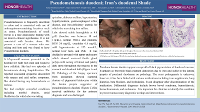
Labs showed stable hemoglobin at 9.8 g/dL (baseline was between 10 and 11g/dL), normal WBCs and platelets, BUN 38 mg/dL, creatinine 0.8 mg/dL, mild hyponatremia at 133 mmol/L, normal liver tests, and INR. It was decided to proceed with upper endoscopy which showed scattered fundic gland polyps with oozing of blood, and patchy dark spots throughout the mucosa in the stomach and duodenal bulb (Figure A and B). Pathology of the biopsy specimen from duodenum showed scattered clusters of pigmented histiocytes within the lamina propria suggestive of pseudomelanosis duodeni (Figure C). She received proton pump inhibitor and was managed with antibiotics for her primary diagnosis of cellulitis and was discharged.
Discussion: Pseudomelanosis duodeni appears as speckled black pigmentation of duodenal mucosa. It appears as brownish black pigment deposition due to iron and sulfur in the lamina propria of proximal duodenum on pathology. The exact pathogenesis is unknown; however, it has been linked with various medications including iron supplements, loop diuretics, beta blockers, and hydralazine. It can disappear after discontinuation of the medication. Differential diagnoses include brown bowel syndrome, hemosiderosis, hemochromatosis, and melanoma. It is important for clinician to identify this condition to prevent unnecessary diagnostic workup and interventions.

Disclosures:
Faisal Mehmood, MD1, Hajra Jamil, MD2, Joseph Fares, MD3, Alexander T. Lee, MD3, Christopher Stasik, DO1, Gavin Levinthal, MD3. P3272 - Pseudomelanosis Duodeni: Iron’s Duodenal Shade, ACG 2024 Annual Scientific Meeting Abstracts. Philadelphia, PA: American College of Gastroenterology.
1HonorHealth, Phoenix, AZ; 2Services Institute of Medical Sciences, Lahore, Punjab, Pakistan; 3HonorHealth, Scottsdale, AZ
Introduction: Pseudomelanosis is frequently described in colon and is associated with use of anthraquinone-containing laxatives such as senna. Pseudomelanosis of small bowel is a rare endoscopic finding with no known clinical significance. It is not associated with laxative abuse. We present a case of a woman who was taking oral iron and was found to have Pseudomonas duodeni.
Case Description/Methods: A 65-year-old woman presented to the hospital for right foot pain and found to have cellulitis. She had two episodes of hematemesis during hospitalization. She reported associated epigastric discomfort with nausea and acid reflux symptoms. She denied having any hematochezia or melena.
She had multiple comorbid conditions including morbid obesity, atrial fibrillation for which she was taking apixaban, diabetes mellitus, hypertension, hypothyroidism, gastroesophageal reflux disease, and iron-deficiency anemia for which she was taking iron sulfate.
Labs showed stable hemoglobin at 9.8 g/dL (baseline was between 10 and 11g/dL), normal WBCs and platelets, BUN 38 mg/dL, creatinine 0.8 mg/dL, mild hyponatremia at 133 mmol/L, normal liver tests, and INR. It was decided to proceed with upper endoscopy which showed scattered fundic gland polyps with oozing of blood, and patchy dark spots throughout the mucosa in the stomach and duodenal bulb (Figure A and B). Pathology of the biopsy specimen from duodenum showed scattered clusters of pigmented histiocytes within the lamina propria suggestive of pseudomelanosis duodeni (Figure C). She received proton pump inhibitor and was managed with antibiotics for her primary diagnosis of cellulitis and was discharged.
Discussion: Pseudomelanosis duodeni appears as speckled black pigmentation of duodenal mucosa. It appears as brownish black pigment deposition due to iron and sulfur in the lamina propria of proximal duodenum on pathology. The exact pathogenesis is unknown; however, it has been linked with various medications including iron supplements, loop diuretics, beta blockers, and hydralazine. It can disappear after discontinuation of the medication. Differential diagnoses include brown bowel syndrome, hemosiderosis, hemochromatosis, and melanoma. It is important for clinician to identify this condition to prevent unnecessary diagnostic workup and interventions.

Figure: (A) Duodenal bulb with patchy dark spots throughout the mucosa in the stomach and duodenal bulb (B) Retroflex view of stomach with oozing gastric polyps (C) High power field image of H & E showing benign duodenal mucosa containing pigmented histiocytes within the lamina propria.
Disclosures:
Faisal Mehmood indicated no relevant financial relationships.
Hajra Jamil indicated no relevant financial relationships.
Joseph Fares indicated no relevant financial relationships.
Alexander Lee indicated no relevant financial relationships.
Christopher Stasik indicated no relevant financial relationships.
Gavin Levinthal indicated no relevant financial relationships.
Faisal Mehmood, MD1, Hajra Jamil, MD2, Joseph Fares, MD3, Alexander T. Lee, MD3, Christopher Stasik, DO1, Gavin Levinthal, MD3. P3272 - Pseudomelanosis Duodeni: Iron’s Duodenal Shade, ACG 2024 Annual Scientific Meeting Abstracts. Philadelphia, PA: American College of Gastroenterology.
