Monday Poster Session
Category: General Endoscopy
P2441 - Minimally Invasive Diagnosis of Linitis Plastica With EUS-FNA
Monday, October 28, 2024
10:30 AM - 4:00 PM ET
Location: Exhibit Hall E

Has Audio

Rohan Rau, DO
Albert Einstein Medical Center
Philadelphia, PA
Presenting Author(s)
Rohan Rau, DO1, Edward J. Marines, DO2, Priya Varghese, MD1, Michael Goldberg, DO1, Maria Lagarde Mussa, MD3
1Albert Einstein Medical Center, Philadelphia, PA; 2Jefferson Einstein Hospital, Philadelphia, PA; 3Einstein Medical Center, Philadelphia, PA
Introduction: Linitis Plastica (LP) is a subtype of gastric adenocarcinoma characterized by diffuse infiltration into the submucosa and muscularis propria. Early diagnosis can elude clinicians as biopsies of the mucosa fail to penetrate to the affected area. Thus, patients may require an invasive approach such as exploratory laparotomy for a full thickness biopsy. We present a case of linitis plastica which was successfully diagnosed in a minimally invasive approach via fine needle aspiration (FNA).
Case Description/Methods: A 64-year-old female with a past medical history of hyperlipidemia presented to the gastroenterology office for reflux symptoms. Initial esophagogastroduodenoscopy (EGD) was normal. Follow up EGD demonstrated severe inflammation in the gastric body, antral luminal narrowing and decreased expansion of the gastric lumen on insufflation. Biopsies showed extensive complete intestinal metaplasia.
Computed tomography of the abdomen (CTA) found diffuse wall thickening of the stomach, perigastric free fluid, moderate ascites and a nodular liver. Paracentesis revealed low serum ascites albumin gradient with high protein, consistent with a non-portal hypertensive etiology. Paracentesis fluid was negative for malignant cells.
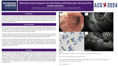
Endoscopic ultrasound (EUS) demonstrated diffuse wall thickening in the gastric body. Layer 4 was thickened at 5 millimeters and the gastric wall was 12 millimeters. FNA biopsy successfully targeted the muscularis propria with four passes of a 22-gauge ultrasound core biopsy needle. Histology showed poorly differentiated adenocarcinoma, therefore avoiding surgery for a full thickness biopsy. She established care with oncology and initiated treatment with FOLFOX.
Discussion: Diagnosis of mucosal gastric adenocarcinomas can be accomplished via jumbo forceps. Submucosal malignancies such as linitis plastica are challenging to biopsy as mucosa appears normal and biopsies rarely sample the submucosa. The “Strip and bite'' technique, “bite-on-bite” technique, or full thickness biopsy can provide submucosal biopsies. Full thickness biopsies via exploratory laparotomy leads to increased morbidity thus minimally invasive techniques are preferred. Our case highlights the diagnostic difficulty with traditional EGD biopsies, advantages of EUS in identifying focal areas of concern and the benefit of FNA to provide an accurate diagnosis. EUS guided FNA as conducted here can provide a minimally invasive alternative that successfully obtains submucosal tissue.

Disclosures:
Rohan Rau, DO1, Edward J. Marines, DO2, Priya Varghese, MD1, Michael Goldberg, DO1, Maria Lagarde Mussa, MD3. P2441 - Minimally Invasive Diagnosis of Linitis Plastica With EUS-FNA, ACG 2024 Annual Scientific Meeting Abstracts. Philadelphia, PA: American College of Gastroenterology.
1Albert Einstein Medical Center, Philadelphia, PA; 2Jefferson Einstein Hospital, Philadelphia, PA; 3Einstein Medical Center, Philadelphia, PA
Introduction: Linitis Plastica (LP) is a subtype of gastric adenocarcinoma characterized by diffuse infiltration into the submucosa and muscularis propria. Early diagnosis can elude clinicians as biopsies of the mucosa fail to penetrate to the affected area. Thus, patients may require an invasive approach such as exploratory laparotomy for a full thickness biopsy. We present a case of linitis plastica which was successfully diagnosed in a minimally invasive approach via fine needle aspiration (FNA).
Case Description/Methods: A 64-year-old female with a past medical history of hyperlipidemia presented to the gastroenterology office for reflux symptoms. Initial esophagogastroduodenoscopy (EGD) was normal. Follow up EGD demonstrated severe inflammation in the gastric body, antral luminal narrowing and decreased expansion of the gastric lumen on insufflation. Biopsies showed extensive complete intestinal metaplasia.
Computed tomography of the abdomen (CTA) found diffuse wall thickening of the stomach, perigastric free fluid, moderate ascites and a nodular liver. Paracentesis revealed low serum ascites albumin gradient with high protein, consistent with a non-portal hypertensive etiology. Paracentesis fluid was negative for malignant cells.
Endoscopic ultrasound (EUS) demonstrated diffuse wall thickening in the gastric body. Layer 4 was thickened at 5 millimeters and the gastric wall was 12 millimeters. FNA biopsy successfully targeted the muscularis propria with four passes of a 22-gauge ultrasound core biopsy needle. Histology showed poorly differentiated adenocarcinoma, therefore avoiding surgery for a full thickness biopsy. She established care with oncology and initiated treatment with FOLFOX.
Discussion: Diagnosis of mucosal gastric adenocarcinomas can be accomplished via jumbo forceps. Submucosal malignancies such as linitis plastica are challenging to biopsy as mucosa appears normal and biopsies rarely sample the submucosa. The “Strip and bite'' technique, “bite-on-bite” technique, or full thickness biopsy can provide submucosal biopsies. Full thickness biopsies via exploratory laparotomy leads to increased morbidity thus minimally invasive techniques are preferred. Our case highlights the diagnostic difficulty with traditional EGD biopsies, advantages of EUS in identifying focal areas of concern and the benefit of FNA to provide an accurate diagnosis. EUS guided FNA as conducted here can provide a minimally invasive alternative that successfully obtains submucosal tissue.

Figure: Figure 1: Edematous and erythematous gastric body which demonstrated poor insufflation. Figure 2: Thickened gastric wall, approximately 12mm. Figure 3: 40x HE stain of malignant ascites fluid showing metastatic high-grade adenocarcinoma. The ascites fluid displays a classic signet ring cell with the nucleus pushed to the periphery. Figure 4: EUS guided FNA.
Disclosures:
Rohan Rau indicated no relevant financial relationships.
Edward Marines indicated no relevant financial relationships.
Priya Varghese indicated no relevant financial relationships.
Michael Goldberg indicated no relevant financial relationships.
Maria Lagarde Mussa indicated no relevant financial relationships.
Rohan Rau, DO1, Edward J. Marines, DO2, Priya Varghese, MD1, Michael Goldberg, DO1, Maria Lagarde Mussa, MD3. P2441 - Minimally Invasive Diagnosis of Linitis Plastica With EUS-FNA, ACG 2024 Annual Scientific Meeting Abstracts. Philadelphia, PA: American College of Gastroenterology.
