Tuesday Poster Session
Category: Small Intestine
P5019 - Carcinoid Tumor in the Ampulla: A Case Report
Tuesday, October 29, 2024
10:30 AM - 4:00 PM ET
Location: Exhibit Hall E

Has Audio

Zhexiang He, DO
Conway Regional Health System
Conway, AR
Presenting Author(s)
Zhexiang He, DO1, Sweehoney Vujjini, MD1, Lauren (Jiyoon) Park, BS, MS2, Sohaib Rana, MD1, Greg Kendrick, MD1, Owen Maat, MD1
1Conway Regional Health System, Conway, AR; 2Conway Regional Health System, Richmond Heights, OH
Introduction: In 1867, Theodor Langhans provided the first histological description of carcinoid tumors. These tumors are believed to originate from enterochromaffin cells and are distributed throughout the entire gastrointestinal system. The annual incidence rate is around 2.5 cases per 100,000.
The small intestine is the most common location, accounting for approximately 44.7% of all cases, followed by the rectum, appendix, and stomach. Carcinoid tumors originating from the ampulla of Vater are rare. In this report, we present a case of a carcinoid tumor originating from the ampulla of Vater.
Case Description/Methods: The patient is a 69-year-old Hispanic woman with PMHs of hypertension, hypothyroidism, and anemia presented at Emergency Department with complaints of abdominal pain, nausea, and vomiting for a few days. The patient mentioned intermittent abdominal pain that started approximately four months ago.
While in the emergency department, the patient had leukocytosis, with a WBC count of 19,000, and an elevated total bilirubin level of 1.3. All liver enzymes were unremarkable.
A CT scan of the abdomen and pelvis showed biliary ductal dilation and evidence of a filling defect located distally in the common bile duct, without liver abnormalities. MRCP reveals mild dilation of the common bile duct, with a maximum diameter of 8 mm.
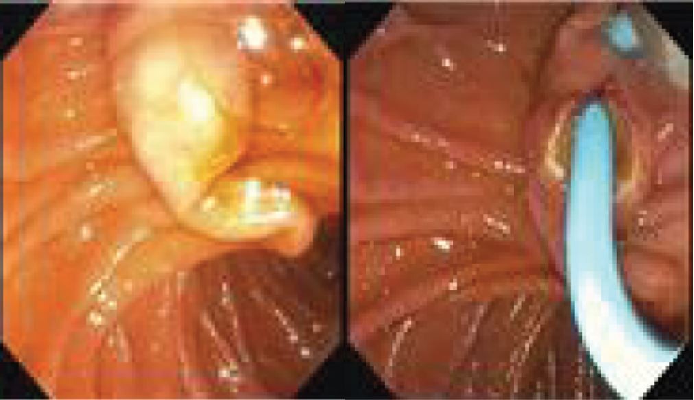
The patient underwent ERCP and a benign-appearing stricture resembling a single-shelf stricture was observed at the ampulla. No obvious signs of adenomatous changes were present, and no pre-obstructive dilation. The cholangiogram showed diffuse dilation of the common hepatic duct and common bile duct, measuring approximately 9 mm in diameter.
The pathology report indicates superficial small bowel mucosa with the presence of a low-grade neuroendocrine carcinoma (NE-1), carcinoid tumor. INSM1 and synaptophysin stain were positive, and the Ki67 proliferation rate was found to be less than 1%.
The patient underwent two more ERCPs for total resection of the carcinoid tumor.
Discussion: Neuroendocrine tumors (NETs) occurring in the ampulla of Vater are exceptionally rare, accounting for less than 1% of all gastrointestinal NETs, with approximately 150 documented cases to date. Our patient was presented with atypical symptoms, including abdominal pain, nausea, and vomiting.
Our patient successfully underwent a complete resection of the low-grade tumor. Despite its rarity, clinicians should maintain clinical awareness of NETs in patients presenting with vague gastrointestinal symptoms.

Disclosures:
Zhexiang He, DO1, Sweehoney Vujjini, MD1, Lauren (Jiyoon) Park, BS, MS2, Sohaib Rana, MD1, Greg Kendrick, MD1, Owen Maat, MD1. P5019 - Carcinoid Tumor in the Ampulla: A Case Report, ACG 2024 Annual Scientific Meeting Abstracts. Philadelphia, PA: American College of Gastroenterology.
1Conway Regional Health System, Conway, AR; 2Conway Regional Health System, Richmond Heights, OH
Introduction: In 1867, Theodor Langhans provided the first histological description of carcinoid tumors. These tumors are believed to originate from enterochromaffin cells and are distributed throughout the entire gastrointestinal system. The annual incidence rate is around 2.5 cases per 100,000.
The small intestine is the most common location, accounting for approximately 44.7% of all cases, followed by the rectum, appendix, and stomach. Carcinoid tumors originating from the ampulla of Vater are rare. In this report, we present a case of a carcinoid tumor originating from the ampulla of Vater.
Case Description/Methods: The patient is a 69-year-old Hispanic woman with PMHs of hypertension, hypothyroidism, and anemia presented at Emergency Department with complaints of abdominal pain, nausea, and vomiting for a few days. The patient mentioned intermittent abdominal pain that started approximately four months ago.
While in the emergency department, the patient had leukocytosis, with a WBC count of 19,000, and an elevated total bilirubin level of 1.3. All liver enzymes were unremarkable.
A CT scan of the abdomen and pelvis showed biliary ductal dilation and evidence of a filling defect located distally in the common bile duct, without liver abnormalities. MRCP reveals mild dilation of the common bile duct, with a maximum diameter of 8 mm.
The patient underwent ERCP and a benign-appearing stricture resembling a single-shelf stricture was observed at the ampulla. No obvious signs of adenomatous changes were present, and no pre-obstructive dilation. The cholangiogram showed diffuse dilation of the common hepatic duct and common bile duct, measuring approximately 9 mm in diameter.
The pathology report indicates superficial small bowel mucosa with the presence of a low-grade neuroendocrine carcinoma (NE-1), carcinoid tumor. INSM1 and synaptophysin stain were positive, and the Ki67 proliferation rate was found to be less than 1%.
The patient underwent two more ERCPs for total resection of the carcinoid tumor.
Discussion: Neuroendocrine tumors (NETs) occurring in the ampulla of Vater are exceptionally rare, accounting for less than 1% of all gastrointestinal NETs, with approximately 150 documented cases to date. Our patient was presented with atypical symptoms, including abdominal pain, nausea, and vomiting.
Our patient successfully underwent a complete resection of the low-grade tumor. Despite its rarity, clinicians should maintain clinical awareness of NETs in patients presenting with vague gastrointestinal symptoms.

Figure: Benign-appearing ampullar stricture that guidewire was able to advance through CBD.
Disclosures:
Zhexiang He indicated no relevant financial relationships.
Sweehoney Vujjini indicated no relevant financial relationships.
Lauren (Jiyoon) Park indicated no relevant financial relationships.
Sohaib Rana indicated no relevant financial relationships.
Greg Kendrick indicated no relevant financial relationships.
Owen Maat indicated no relevant financial relationships.
Zhexiang He, DO1, Sweehoney Vujjini, MD1, Lauren (Jiyoon) Park, BS, MS2, Sohaib Rana, MD1, Greg Kendrick, MD1, Owen Maat, MD1. P5019 - Carcinoid Tumor in the Ampulla: A Case Report, ACG 2024 Annual Scientific Meeting Abstracts. Philadelphia, PA: American College of Gastroenterology.
