Monday Poster Session
Category: Colon
P2010 - An Unexpected Discovery During a Surveillance Colonoscopy: Mucinous Appendiceal Neoplasm
Monday, October 28, 2024
10:30 AM - 4:00 PM ET
Location: Exhibit Hall E

Has Audio
- FM
Faisal Mehmood, MD
HonorHealth
Glendale, AZ
Presenting Author(s)
Faisal Mehmood, MD1, Hajra Jamil, MD2, Bassel AL-Lahham, MD3, Neeraj Singh, MD4, Joseph Fares, MD5, Christopher Stasik, DO1, Gavin Levinthal, MD5
1HonorHealth, Phoenix, AZ; 2Services Institute of Medical Sciences, Lahore, Punjab, Pakistan; 3HonorHealth North Valley Gastroenterology, Phoenix, AZ; 4Colon and Rectal Care Center of Phoenix, Glendale, AZ; 5HonorHealth, Scottsdale, AZ
Introduction: Appendiceal mucocele is a descriptive term for an abnormal mucous accumulation distending the appendiceal lumen. It was first described in 1842 by Rokitansky. These neoplasms can be asymptomatic for many years. We present a case of an asymptomatic middle-aged woman who had a bulging appendiceal orifice on a colonoscopy and later was diagnosed with mucinous appendiceal neoplasm.
Case Description/Methods: A 61-year-old female presented to the ambulatory surgery center for a surveillance colonoscopy. Her past medical history was unremarkable. She was asymptomatic.
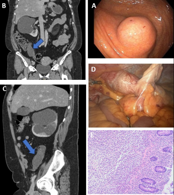
Her blood work including complete blood count and comprehensive metabolic panel were unremarkable. She underwent a colonoscopy and was found to have a bulging appendiceal orifice which was flattening with peristaltic waves (Figure A).
After the colonoscopy, it was decided to get a computerized tomography (CT) of abdomen and pelvis with IV contrast. It was noted that she has an elongated, well-circumscribed low-density lesion in the region of the appendix measuring 6.8 x 3.9 x 3.1 cm and was highly suspicious for mucocele of the appendix (Figure B & C).
Colorectal surgery decided to pursue a surgical resection. Intraoperatively, she was found to have a large appendiceal mucocele and cystic lesion involving the entire appendix and the base of the cecum (Figure D). She underwent a robotic right hemicolectomy with side-to-side anastomosis between distal ascending colon and distal ileum. Postoperative course was uneventful. Pathology showed low-grade mucinous appendiceal neoplasm measuring 9.5 cm (Figure E). Resection margins were negative for malignancy. Nine lymph nodes were removed and were negative for malignancy. She was doing well on a follow-up visit.
Discussion: Appendiceal mucinous neoplasms are extremely rare. The estimated prevalence is less than 0.5% of all GI neoplasms. In the United States, approximately 3500 cases are diagnosed annually. Females are predominantly affected and are diagnosed in the sixth decade of life. These neoplasms are usually an incidental finding during imaging, colonoscopy, or in the pathology specimen of the appendix. However, advanced disease can present with nonspecific symptoms including abdominal pain or palpable mass. Management depends on the staging of the disease. Low-grade tumors are treated surgically with a resection of the primary tumor. However, advanced disease can be managed with peritoneal debulking with chemotherapy.

Disclosures:
Faisal Mehmood, MD1, Hajra Jamil, MD2, Bassel AL-Lahham, MD3, Neeraj Singh, MD4, Joseph Fares, MD5, Christopher Stasik, DO1, Gavin Levinthal, MD5. P2010 - An Unexpected Discovery During a Surveillance Colonoscopy: Mucinous Appendiceal Neoplasm, ACG 2024 Annual Scientific Meeting Abstracts. Philadelphia, PA: American College of Gastroenterology.
1HonorHealth, Phoenix, AZ; 2Services Institute of Medical Sciences, Lahore, Punjab, Pakistan; 3HonorHealth North Valley Gastroenterology, Phoenix, AZ; 4Colon and Rectal Care Center of Phoenix, Glendale, AZ; 5HonorHealth, Scottsdale, AZ
Introduction: Appendiceal mucocele is a descriptive term for an abnormal mucous accumulation distending the appendiceal lumen. It was first described in 1842 by Rokitansky. These neoplasms can be asymptomatic for many years. We present a case of an asymptomatic middle-aged woman who had a bulging appendiceal orifice on a colonoscopy and later was diagnosed with mucinous appendiceal neoplasm.
Case Description/Methods: A 61-year-old female presented to the ambulatory surgery center for a surveillance colonoscopy. Her past medical history was unremarkable. She was asymptomatic.
Her blood work including complete blood count and comprehensive metabolic panel were unremarkable. She underwent a colonoscopy and was found to have a bulging appendiceal orifice which was flattening with peristaltic waves (Figure A).
After the colonoscopy, it was decided to get a computerized tomography (CT) of abdomen and pelvis with IV contrast. It was noted that she has an elongated, well-circumscribed low-density lesion in the region of the appendix measuring 6.8 x 3.9 x 3.1 cm and was highly suspicious for mucocele of the appendix (Figure B & C).
Colorectal surgery decided to pursue a surgical resection. Intraoperatively, she was found to have a large appendiceal mucocele and cystic lesion involving the entire appendix and the base of the cecum (Figure D). She underwent a robotic right hemicolectomy with side-to-side anastomosis between distal ascending colon and distal ileum. Postoperative course was uneventful. Pathology showed low-grade mucinous appendiceal neoplasm measuring 9.5 cm (Figure E). Resection margins were negative for malignancy. Nine lymph nodes were removed and were negative for malignancy. She was doing well on a follow-up visit.
Discussion: Appendiceal mucinous neoplasms are extremely rare. The estimated prevalence is less than 0.5% of all GI neoplasms. In the United States, approximately 3500 cases are diagnosed annually. Females are predominantly affected and are diagnosed in the sixth decade of life. These neoplasms are usually an incidental finding during imaging, colonoscopy, or in the pathology specimen of the appendix. However, advanced disease can present with nonspecific symptoms including abdominal pain or palpable mass. Management depends on the staging of the disease. Low-grade tumors are treated surgically with a resection of the primary tumor. However, advanced disease can be managed with peritoneal debulking with chemotherapy.

Figure: (A) Colonoscopy view of bulging appendiceal orifice
(B & C) Coronal and sagittal view of CT abdomen and pelvis with IV contrast showing an elongated, well-circumscribed low-density lesion in the region of the appendix measuring 6.8 x 3.9 x 3.1 cm
(D) Intraoperative view of appendix with a large appendiceal mucocele and cystic lesion involving the entire appendix and the base of cecum.
(E) High power view of surface epithelium showing mucin producing epithelial cells and no evidence of high grade dysplasia and demonstrates the absence of a muscularis mucosa layer suggestive of low-grade mucinous appendiceal neoplasm
Disclosures:
Faisal Mehmood indicated no relevant financial relationships.
Hajra Jamil indicated no relevant financial relationships.
Bassel AL-Lahham indicated no relevant financial relationships.
Neeraj Singh indicated no relevant financial relationships.
Joseph Fares indicated no relevant financial relationships.
Christopher Stasik indicated no relevant financial relationships.
Gavin Levinthal indicated no relevant financial relationships.
Faisal Mehmood, MD1, Hajra Jamil, MD2, Bassel AL-Lahham, MD3, Neeraj Singh, MD4, Joseph Fares, MD5, Christopher Stasik, DO1, Gavin Levinthal, MD5. P2010 - An Unexpected Discovery During a Surveillance Colonoscopy: Mucinous Appendiceal Neoplasm, ACG 2024 Annual Scientific Meeting Abstracts. Philadelphia, PA: American College of Gastroenterology.
